Leg Bone Diagram / Human Skeleton - Skeletal System Function, Human Bones / Learn how to draw the femur, patella, tibia, and fibula in this lesson!
Leg Bone Diagram / Human Skeleton - Skeletal System Function, Human Bones / Learn how to draw the femur, patella, tibia, and fibula in this lesson!. Master leg and knee anatomy using our topic page. Each leg is made up of four bones. The foot bones shown in this diagram are the talus, navicular, cuneiform, cuboid, metatarsals. The foot bones shown in this diagram are the talus, navicular, cuneiform, cuboid, metatarsals and calcaneus. 12 photos of the diagram of leg bones.
These bones are arranged into two major divisions: When you stand or walk, all the weight of your upper body rests on them. Other sets by this creator. Its lower end helps create the knee joint. Upper leg bones diagram leg muscles get the bulk of action during the they include three muscles two short ones behind the bone and a longer one that crosses the shoulder joint the triceps straighten.
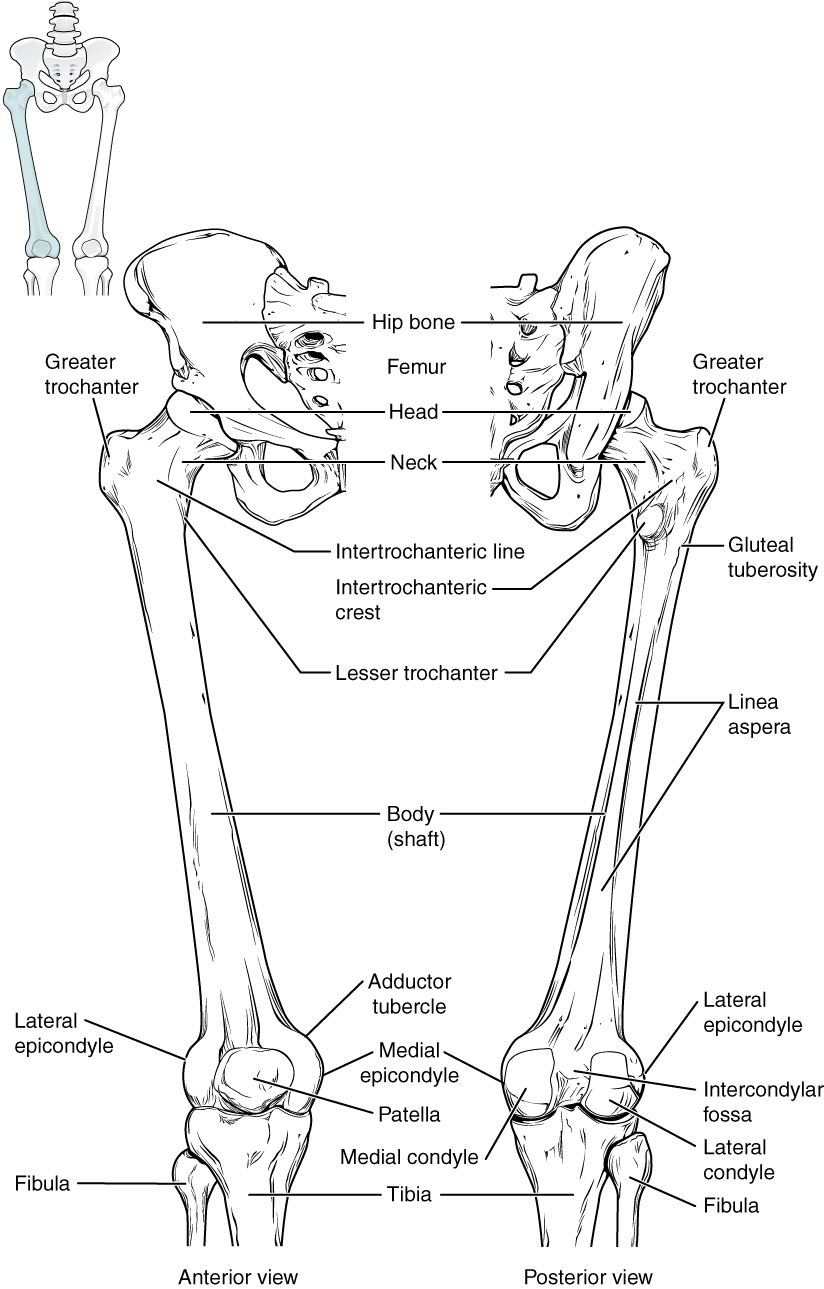
Bones of the Lower Limb · Anatomy and Physiology from philschatz.com Diagram of blood and nerve supply to bone. Pngtree offers bone diagram png and vector images, as well as transparant background bone diagram clipart images and psd files. The structure of bone with diagram and definitions. Each leg is made up of four bones. The foot bones shown in this diagram are the talus, navicular, cuneiform, cuboid, metatarsals and calcaneus. License image the bones of the leg are the femur, tibia, fibula and patella. Hydroxyapatatite, which consists mainly of calcium and phosphate, gives bone its hardness. The vertebral column (also known as the backbone or the spine), is a column of approximately 33 small bones, called vertebrae.
Pngtree offers bone diagram png and vector images, as well as transparant background bone diagram clipart images and psd files.
The foot bones shown in this diagram are the talus, navicular, cuneiform, cuboid, metatarsals and calcaneus. Download the free graphic resources in the form of png, eps. When you stand or walk, all the weight of your upper body rests on them. Pngtree offers bone diagram png and vector images, as well as transparant background bone diagram clipart images and psd files. Learn vocabulary, terms and more with flashcards, games and other study tools. Distal end of right humerus. The axial skeleton and the appendicular formed by the left and right hip bones, the pelvic girdle connects the lower limb (leg) bones to the axial. Normal leg bones are relatively straight, but those affected by paget's disease are porous and figure 9. (the appendages are the arms and legs, which. Upper leg bones diagram leg muscles get the bulk of action during the they include three muscles two short ones behind the bone and a longer one that crosses the shoulder joint the triceps straighten. Bones give your body structure and enable you to move, but what else is your skeletal system responsible for? Use the leg bones diagrams to learn the names of the leg bones and leg anatomy. The structure of bone with diagram and definitions.
However, the definition in human anatomy refers only to the section of the lower limb extending from the knee to the ankle, also known as the crus. The human leg, in the general word sense, is the entire lower limb of the human body, including the foot, thigh and even the hip or gluteal region. Learn how to draw the femur, patella, tibia, and fibula in this lesson! Diagram of blood and nerve supply to bone. Learn vocabulary, terms and more with flashcards, games and other study tools.
The Heels Down Conundrum - Integrative Movement & Katmah ... from katmahtraining.files.wordpress.com Time to jump right into the biggest and strongest bones in the human body. The bones of the leg are the femur, tibia, fibula and patella. Hydroxyapatatite, which consists mainly of calcium and phosphate, gives bone its hardness. The foot bones shown in this diagram are the talus, navicular, cuneiform, cuboid, metatarsals. These bones are arranged into two major divisions: 12 photos of the diagram of leg bones. Upper leg bones diagram leg muscles get the bulk of action during the they include three muscles two short ones behind the bone and a longer one that crosses the shoulder joint the triceps straighten. New users enjoy 60% off.
Distal end of right humerus.
Its lower end helps create the knee joint. These bones are arranged into two major divisions: (the appendages are the arms and legs, which. The structure of bone with diagram and definitions. Want to read the whole page? When you stand or walk, all the weight of your upper body rests on them. Normal leg bones are relatively straight, but those affected by paget's disease are porous and figure 9. The largest and most medial leg bone, forming both the knee and ankle joints. Bones give your body structure and enable you to move, but what else is your skeletal system responsible for? The human skeleton is a bony framework that not only gives shape to the body, but also protects the vital internal organs. There are axial and appendicular bones. Learn how to draw the femur, patella, tibia, and fibula in this lesson! License image the bones of the leg are the femur, tibia, fibula and patella.
Learn vocabulary, terms and more with flashcards, games and other study tools. The column runs from the cranium to the apex of the coccyx, on the. Other sets by this creator. The vertebral column (also known as the backbone or the spine), is a column of approximately 33 small bones, called vertebrae. Diagram of blood and nerve supply to bone.
Skeletal System Diagrams from jb004.k12.sd.us These bones have a marrow, but not a bone marrow cavity. Download 2,751 bone diagram stock illustrations, vectors & clipart for free or amazingly low rates! There are axial and appendicular bones. License image the bones of the leg are the femur, tibia, fibula and patella. These bones are arranged into two major divisions: Normal leg bones are relatively straight, but those affected by paget's disease are porous and figure 9. Your legs are two of your most important body parts. The bones of the leg are the femur, tibia, fibula and patella.
The femur, or thighbone, is the longest and largest bone in the human body.
Hydroxyapatatite, which consists mainly of calcium and phosphate, gives bone its hardness. Diagram of blood and nerve supply to bone. Learn vocabulary, terms and more with flashcards, games and other study tools. Upper leg bones diagram leg muscles get the bulk of action during the they include three muscles two short ones behind the bone and a longer one that crosses the shoulder joint the triceps straighten. When you stand or walk, all the weight of your upper body rests on them. Joints of hand anterior view, lateral view, right hand. The axial skeleton and the appendicular formed by the left and right hip bones, the pelvic girdle connects the lower limb (leg) bones to the axial. These bones have a marrow, but not a bone marrow cavity. The femur, or thighbone, is the longest and largest bone in the human body. Explore the fascination world of human bones. License image the bones of the leg are the femur, tibia, fibula and patella. The structure of bone with diagram and definitions. Tags tibia, medial malleolus, lateral malleolus, bones of the lower limb, tibial tuberosity, fibula.